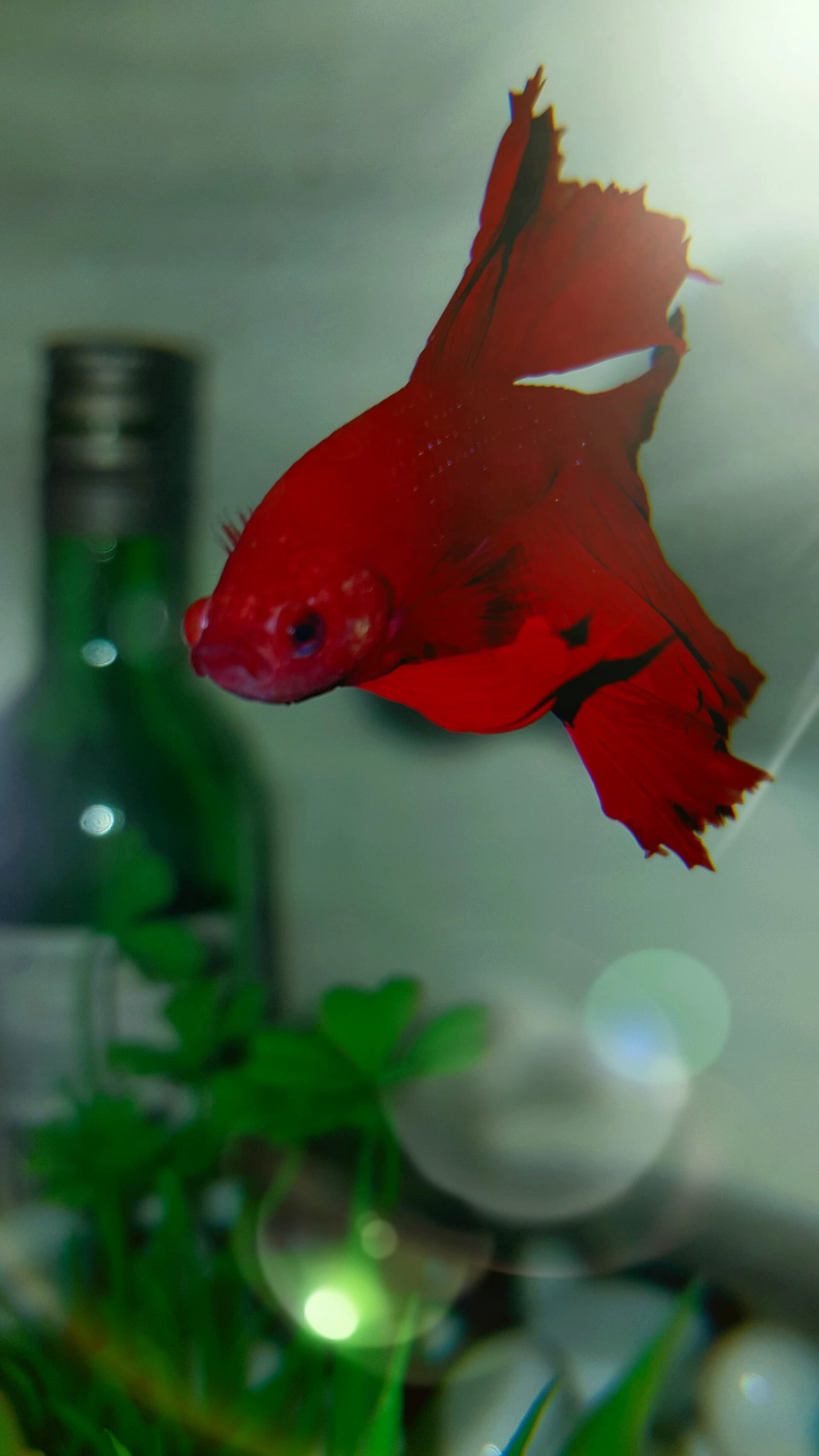
Red streaks on a betta that were not present yesterday are a medical emergency. They mean bacteria are in the bloodstream.
Aeromonas hydrophila is the most common causative organism, though Pseudomonas and, less commonly, Vibrio species can produce the same picture. Veterinary literature groups this presentation under “motile Aeromonas septicemia” (MAS), a name that covers the whole cluster of mobile, gram-negative rods responsible for it, not one specific bug. It is an opportunistic bacterium present in virtually all freshwater aquarium environments at low levels (Merck Veterinary Manual, Bacterial Diseases of Fish). In clean, well-maintained water with a healthy fish, it causes no disease. In a fish compromised by poor water quality, chronic stress, fin rot that was not treated, or an injury that broke the skin barrier, Aeromonas can enter the circulatory system and cause systemic infection (septicemia).
None of these bacteria spread from a sick fish to a healthy one on contact the way a primary pathogen would. They’re opportunists already living in the water column and gut flora of every fish in the tank, sick or not, so there’s no need to treat the whole tank prophylactically the way you might for a genuinely contagious ich outbreak. What made one fish septicemic was that fish’s individual compromise, not exposure. But check the water anyway: if the underlying cause is tank-wide, a failed heater, an ammonia spike, an aggressive tankmate inflicting repeated fin damage, other fish sharing that water are at elevated risk from the same failure, not from contagion. That’s also why time matters so much once streaking appears: kanamycin or NeoPlex started within the first 24 to 48 hours of visible symptoms has a reasonable survival rate. Wait past that window into obvious lethargy, extensive hemorrhaging, and pale gills, and the odds drop sharply.
What it looks like
Early stage:
- Single or multiple red streaks running along fin rays or through the tail fin
- Red spots (petechiae): small pinpoint hemorrhages on the body surface
- Increased gill movement (respiratory distress)
- Fish still active and eating
Progressing:
- Streaks spreading and darkening
- Body pallor in areas around lesions
- Lethargy, reduced feeding
- Possible hemorrhaging at the base of fins (the fin-body junction turns dark red)
Late stage:
- Extensive hemorrhaging across body and all fins
- Pale or grey gills (indicates anemia from blood cell destruction)
- Fish immobile or barely swimming
- Skin may show ulceration at surface lesion sites
Differential: what else looks like this
| Appearance | More likely cause |
|---|---|
| Red streaks on fin edges only, fin margin ragged | Advanced fin rot progressing toward body |
| Single red spot on body, scales raised nearby | Ulcer/bacterial abscess |
| Red streaks + pine-cone scale profile | Dropsy + septicemia co-occurring (poor prognosis) |
| Red color to skin (not streaks), no fish behavior change | May be natural coloration pattern; compare to baseline photos |
| Red streaks after physical trauma (net, decoration) | Mechanical damage to capillaries; monitor for 24h before treating |
How it’s actually diagnosed
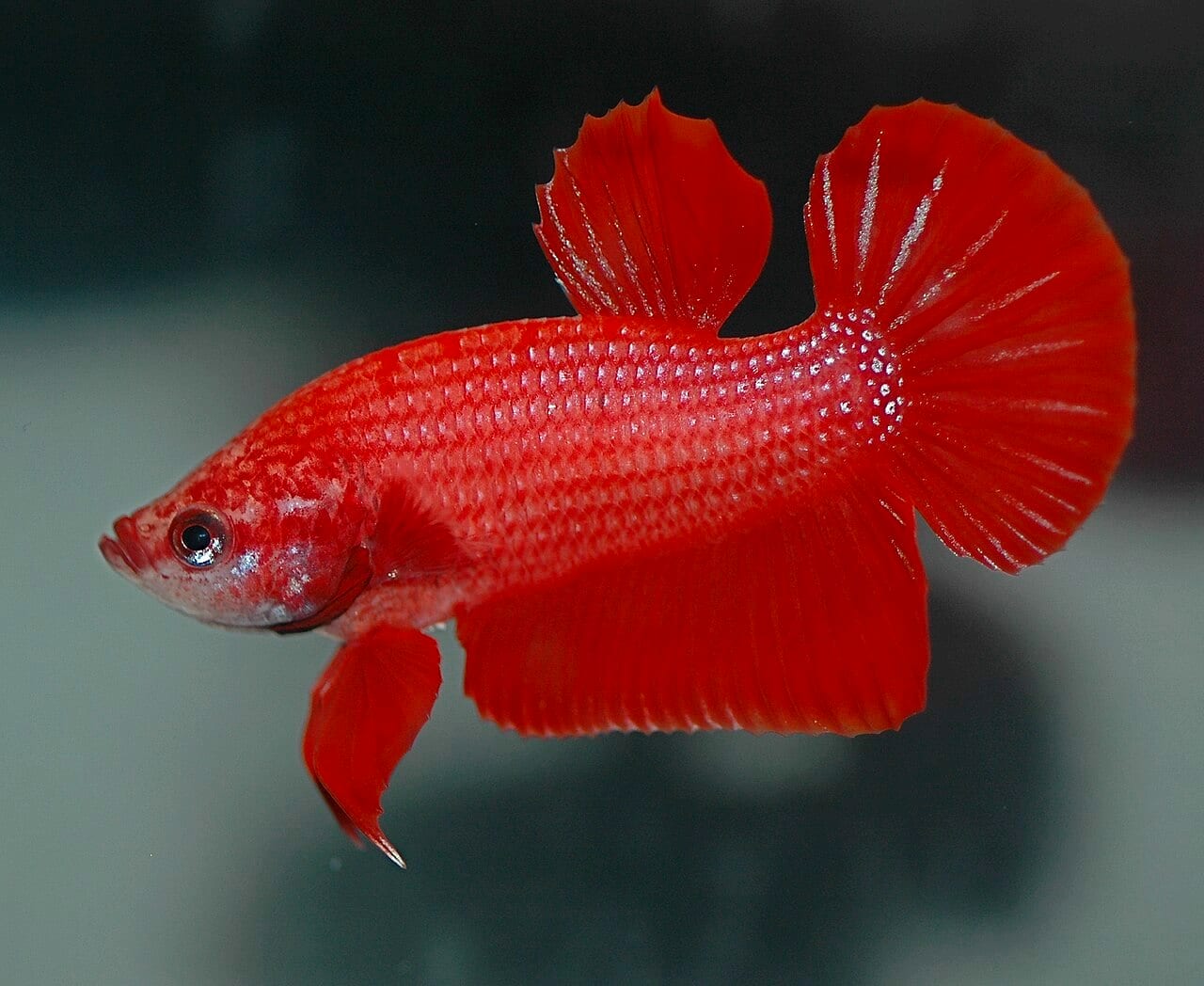
There’s no home test for septicemia. What you’re diagnosing from the couch isn’t a specific organism, it’s a clinical picture. A real diagnosis identifies the causative species and tests it against a panel of antibiotics: a veterinary lab cultures blood or tissue and runs sensitivity testing to see which drugs the strain actually responds to, and blood culture is the preferred method precisely because it’s nonlethal (Merck Veterinary Manual, Bacterial Diseases of Fish). Almost no home aquarist has access to that pipeline, and even where a fish-competent vet exists, culture results take days, longer than the treatment window this disease allows. Septicemia treatment for a pet betta is necessarily empirical: you treat for the organisms proven common in freshwater aquaria, gram-negative and responsive to kanamycin-class drugs, rather than waiting to confirm the exact one.
That empirical approach is why the visible streaking gets treated as an emergency, not a cosmetic issue. Internally, advanced Aeromonas septicemia produces liquefactive necrosis in the spleen and kidney, the organs responsible for a fish’s immune response and blood filtration. Once those are compromised, antibiotic exposure in the water or food can’t reach the infection fast enough to reverse the damage. The streaks you can see are the edge of a process that’s already well underway on the inside.
Treatment protocol
Hospital tank setup
Move the fish immediately. Do not treat the display tank unless other fish are showing symptoms. Bare-bottom 5-gallon tank, pre-seeded sponge filter or air stone, heater at 78-80°F. No substrate: bare bottom allows monitoring of waste and uneaten food.
Antibiotic: first-line
Kanamycin sulfate (Seachem KanaPlex) is the first-line choice for gram-negative septicemia. It is effective against Aeromonas and Pseudomonas and can be dosed in the water or in food. Dosing in food with a gel carrier (Seachem Focus as a binder) delivers the antibiotic directly to internal tissue, which is particularly important for systemic infection.
Follow the KanaPlex dosing instructions for the tank-water method if food-dosing is not practical because the fish has stopped eating.
Seachem KanaPlex on Amazon Affiliate link. See our disclosure.
Seachem NeoPlex (neomycin) is an alternative with good gram-negative coverage, available at most fish stores (Noga, Fish Disease: Diagnosis and Treatment, 2nd ed., 2010).
Why the antibiotic choice isn’t a guarantee
Treat Aeromonas resistance as documented, not hypothetical. A 2021 survey of 161 Aeromonas isolates from diseased ornamental fish and their farm water in Sri Lanka tested 13 antimicrobials and found 51.6% multidrug-resistant. Quinolones, usually the first class reached for against Aeromonas, showed 67.1% resistance in the same isolates. Aminoglycosides, kanamycin’s class, held up better: gentamicin resistance was only 16.8% (Dhanapala et al., Antimicrobial Resistance of Aeromonas from Ornamental Fish, Microorganisms, 2021), part of why kanamycin remains a sound starting choice.
A fish with no improvement after 48 hours on kanamycin may be carrying a resistant strain; switching to NeoPlex is a legitimate next step, not a lost cause. It’s also another argument for finishing the full course: a resistant subpopulation that survives an interrupted course is the selection pressure that breeds more resistance in the tank.
Duration
Treat for a minimum of 10 days. Do not stop because improvement is visible after 5 days; the infection may not be cleared systemically. A shortened antibiotic course is a primary cause of recurrence and antibiotic resistance.
Water changes
30% daily water change with pre-dechlorinated water at the correct temperature, redosing antibiotic proportionally after each change. Daily changes prevent ammonia accumulation in a hospital tank and maintain the water quality that the biological filter of a large display tank would provide.
When treatment isn’t the answer
Start treatment while the fish is still active and responsive; that’s when the odds favor recovery. Be honest with yourself about what “late-stage” looks like, because this disease does not reliably respond to a heroic second or third attempt once it gets there. A fish that’s immobile, hemorrhaging extensively, with gills pale rather than red, has organ damage antibiotics can’t undo even if they finally clear the bacteria. Prognosis at that point is poor regardless of drug or dose, and continuing to medicate a fish that’s stopped eating and can’t right itself in the water is prolonging suffering, not treating a survivable illness. That’s a call for the euthanasia protocol, not more medication.
After recovery
A fish that survives septicemia has been through significant immunological stress. After completing the antibiotic course:
- Do not return to the display tank immediately; keep in the hospital tank for 5–7 more days observing for recurrence
- Do a thorough water change and parameter check of the display tank before return
- Identify and correct the predisposing factor (what allowed Aeromonas to become pathogenic)
The predisposing factor is almost always water quality. See water chemistry and the nitrogen cycle.
Preventing progression from fin rot
Septicemia often follows untreated or under-treated fin rot. Fin rot allows bacteria to breach the tissue boundary. If fin rot is present and progressing, the antibiotic that treats septicemia is the same antibiotic that treats moderate-to-severe fin rot. Beginning treatment before systemic infection develops is the correct approach.